Diagnosis and treatment methods and technologies continue to evolve in the delivery of high-quality oral health care. Like other medical specialties, precision in diagnosis is a cornerstone of effective dental treatment. A major development in diagnostic improvements has been the introduction of advanced imaging technologies. These have significantly transformed the ability of dentists to view dental structures with superior clarity and detail. Among these innovations, Cone Beam Computed Tomography (CBCT) has emerged as a game-changer, setting a new standard for accuracy and comprehensive assessment in dental care.
CBCT technology represents a significant advancement from traditional dental X-rays by offering three-dimensional images that provide a lifelike view of the mouth and surrounding areas. This sophisticated imaging technique not only captures the intricate details of dental anatomy but can also reveal various dental diseases and hidden anomalies–conditions that conventional methods might miss. As a diagnostic and treatment planning tool in modern dentistry, CBCT is revolutionizing multiple aspects of dental practice, from implantology and orthodontics to endodontics and oral surgery.
Understanding CBCT Technology
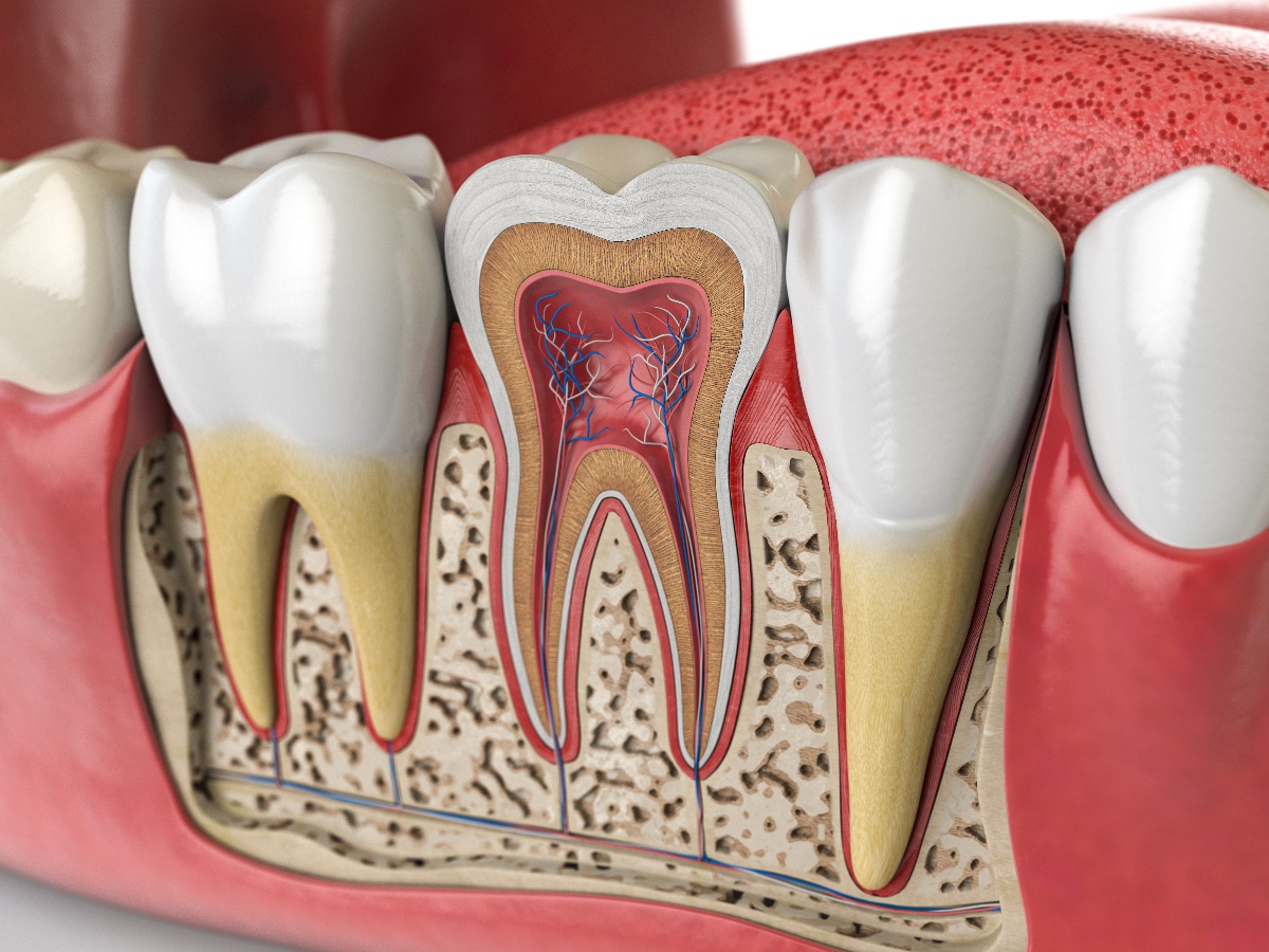
Cone Beam Computed Tomography significantly differs from traditional dental X-rays by providing three-dimensional images that offer a more complete view of the anatomy of your mouth, teeth, jaws, and surrounding areas. As you gain an understanding of how CBCT works, you can appreciate its role in helping your dentist more accurately diagnose and successfully treat dental conditions.
How CBCT Works. A dental CBCT scanner rotates around your head and captures data using a cone-shaped X-ray beam. These images are then reconstructed into a three-dimensional representation of your teeth, jaws, and surrounding areas, including your airway. This process allows your dentist to view your anatomical structures from different angles and planes, which is not possible with standard two-dimensional X-rays.
Differences Between CBCT and Traditional Dental X-Rays
CBCT images differ from traditional X-rays in several key ways, such as:
- Dimensional Imaging. Traditional X-rays provide two-dimensional images, while CBCT offers three-dimensional imaging.
- Accuracy. CBCT images have higher accuracy and detail, revealing structures that might be obscured in two-dimensional X-rays.
- Radiation Exposure. While CBCT involves more radiation than standard dental X-rays, it is typically less than a conventional medical CT scan.
- Field of View. CBCT can be adjusted to scan specific areas (small, medium, or large. This makes it more versatile for different dental procedures.
Advancements in CBCT Technology
Dental technologies such as artificial intelligence (AI) and CBCT continue to transform and improve the delivery of dental care. Some of the advancements in CBCT include:
- Image Quality. Recent advancements have significantly improved the image quality, allowing for clearer and more precise visualizations.
- Reduced Radiation. Newer models have enhanced technology to minimize radiation exposure while maintaining image quality.
- Software Integration. Integration with dental planning software has enabled more accurate treatment planning and simulations.
- Speed and Comfort. Modern CBCT scanners are faster and designed for greater patient comfort during the scanning process.
The Importance of Accurate Diagnosis in Dental Treatment
Accurate diagnosis is the foundation of effective dental treatment because it helps determine the course of action for a dental condition and significantly influences the success of that treatment. Historically, dentists have relied on visual examination and traditional two-dimensional X-rays. While these remain very important for dentists, they come with certain limitations that can affect the precision of diagnosis.
Limitations of Traditional Diagnostic Methods
- Limited Visibility. Traditional X-rays provide only a two-dimensional view, which can obscure certain anatomical structures and oral diseases and conditions.
- Risk of Misdiagnosis. Without comprehensive, lifelike imaging, there's a higher risk of overlooking some issues. This can lead to misdiagnosis or inaccurate treatment planning.
- Difficulty in Detecting Subtle Anomalies. Early stages of certain dental conditions, like hairline fractures or small lesions in the jawbone, might not be visible in 2D images.
Role of CBCT in Overcoming Limitations
- Comprehensive 3D Imaging. CBCT provides three-dimensional images, offering a complete view of the head and neck areas. This reduces the risk of a diagnostic oversight.
- Detailed Visualization. CBCT captures fine details that are critical for diagnosing complex dental issues, such as the extent of periodontal disease, root canal anomalies, and the precise location of impacted teeth.
- Improved Treatment Planning. The accuracy of CBCT imaging aids in creating more effective treatment plans, especially in the placement of dental implants.
- Early Detection. CBCT's superior imaging capabilities enable the early detection of dental problems, potentially leading to less invasive and more successful treatments.
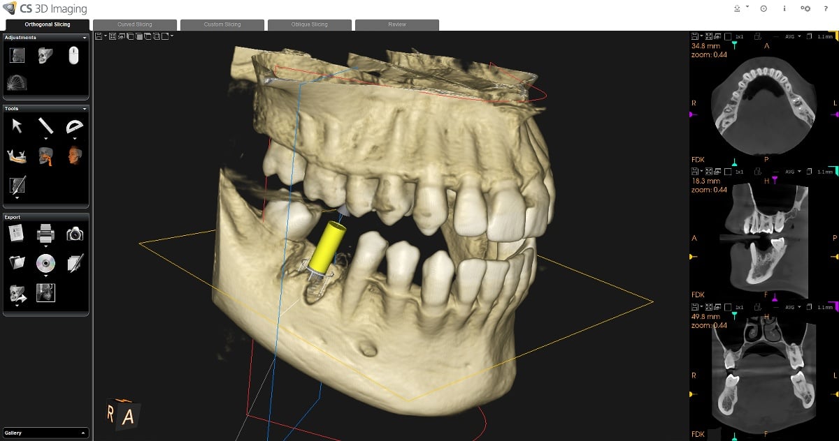
CBCT in the Placement of Dental Implants
In recent years, dental implants have revolutionized the replacement of missing teeth. One of the significant factors in the success of dental implants has been the use of CBCT to more accurately view the surrounding anatomy and the precise place of each implant. This precise placement directly influences the implant's functionality, longevity, and esthetic outcome of each implant and attached restoration.
The key advantages of using CBCT for dental implant placement include:
- Accurate Assessment of Bone Quality and Quantity. CBCT scans allow your dentist to evaluate the density and volume of your jawbone. This is crucial for determining the feasibility of implant placement and if you will need bone grafting.
- Precise Implant Positioning. The detailed imagery helps in planning the implant's exact position, angle, and depth, reducing the risk of damaging vital structures like nerves and sinuses.
- Customized Treatment Planning. CBCT data can be integrated with dental planning software to create a customized surgical guide, ensuring implants are placed precisely as planned.
- Early Detection of Potential Complications. CBCT helps identify any anatomical challenges or potential complications before the surgery, allowing for preemptive adjustments in the treatment plan.
- Improved Patient Safety. The accuracy of CBCT imaging minimizes the risk of intraoperative complications, enhancing overall patient safety.
The importance of CBCT in dental implantology cannot be overstated. It provides a roadmap for the entire implant procedure, from initial consultation to the surgical phase. For instance, CBCT imaging can identify the most suitable sites for implant placement in cases where the jawbone is narrow or has irregularities. This helps ensure the optimal esthetic alignment with the rest of your teeth.
CBCT imaging is especially valuable in complex cases, such as when implants are placed in the posterior upper jaw, a region close to the sinus cavities. Here, the precision of CBCT imaging is instrumental in avoiding sinus perforation. Additionally, for full-arch restorations requiring multiple implants, CBCT ensures that each implant is optimally positioned to support the prosthetic framework effectively.
Schedule an Appointment
To learn more about how Palmetto Dental Arts can use 3D Cat Scan technology to help you improve your smile, call us or contact us online.